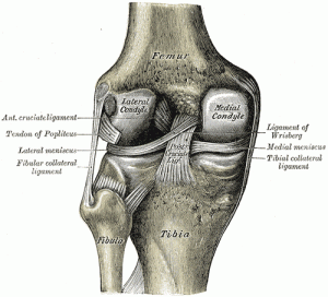
The proximal and distal tibiofibular joints refer to two articulations between the tibia and fibula of the leg. These joints have minimal function in terms of movement but play a greater role in stability and weight-bearing. Some studies show that manual movement of the distal tibiofibular joint led to a significant increase in the range of ankle dorsiflexion.
The articulations between the tibia and fibula are effected by ligaments which connect the extremities and bodies of the bones. The ligaments may consequently be subdivided into three sets:
- those of the Tibiofibular articulation;
- the interosseous membrane;
- those of the Tibiofibular syndesmosis.
Proximal tibiofibular articulation
The proximal tibiofibular articulation (also called superior tibiofibular joint) is an Plane Joint – Arthrodial Joint between the lateral condyle of the tibia and the head of the fibula.
The contiguous surfaces of the bones present flat, oval facets covered with cartilage and connected together by an articular capsule and by anterior and posterior ligaments.
Superior Tibiofibular Joint functions to
- reduce rotational stress
- prevent lateral bending of the tibia
- spread axial loads when standing
Interosseus membrane
Interossoeus membrane provides additional surfaces for attachment of muscles and binds the tibia and the fibula together. It also resists the downward pull exerted on the fibula by the powerful muscles attached to the bone.
Distal tibiofibular articulation
It is a fibrous articulation, a Syndesmosis. Its major features are:
- Inferior tibiofibular joint permits slight movements, so that the lateral malleolus can rotate laterally during dorsiflexion of the ankle.
- The strength of the ligaments of inferior tibiofibular joint is an important factor in maintaining the integrity of the ankle joint.
There are no muscle directly associated with these joints.

